Breast Pathology
What is breast cancer?
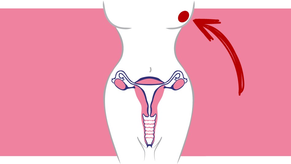
The breast consists of a series of mammary glands interconnected by tubes known as mammary ducts, which carry milk to the nipple during lactation. The mammary glands and ducts are embedded in adipose tissue and the conjunctive tissue, which together with the lymphatic tissue make up the breast.Breast cancer is the abnormal and disordered growth of the cells of this tissue.
Most tumours occurring in the breast are benign tumours, which are non-cancerous and are caused by fibrocystic nodules. The fluid they contain can be drained off and any pain relieved. Benign tumours are largely related to genetic factors. Their symptoms are pain and inflammation, but they do not spread to other parts of the body and are not dangerous.
There are however malign tumours, which are either localized or have spread through the blood or lymphatic vessels, giving rise to metastasis, a cancer in an organ different from that where they originated. Of all the cases of breast cancer, only from 7% to 10% present metastasis at the outset.
15,000 new cases are diagnosed in Spain every year, and one in every 16 or 18 Spanish women are expected to suffer from breast cancer. Breast cancer is the primary cause of death from cancer in women between the ages of 45-55 years, while the the rate of cure in Spain is approximately 60%. Early detection, before the tumour has had the chance to spread, brings the rate of recovery up to almost 90%.
What types of breast cancer are there?
Prognosis and treatment of cancer depends on the extent of its development and the risk factors pertaining to each woman. In order for these to be determined, it is necessary to perform a series of analyses to provide a classification of these conditions. The American Joint Committee on Cancer employs the TNM classification that respectively describes the size and extent of the tumour, as well as whether the cancer has spread to nearby lymph nodes or to other parts of the body.
Classification of the different stages is from I to IV:
I: when the tumour is less than 2 cm and there is no metastasis.
II: when the tumour is less than 2 cm but the axillary lymph nodes are affected, or when it measures from 2-5cm but may not have spread. Also when it is larger than 5cm but regional nodes are not affected.
III: in case A, the tumour measures less than 5cm and has spread to regional nodes or extended to lymph nodes inside the chest wall. In case B, the cancer has spread to other neighbouring tissue or to nodes inside the chest wall, close to the sternum.
IV: it has spread to other structures, such as bones, lungs, the liver or the brain.
We differentiate between different types of breast cancer:
Ductal carcinoma in situ originating in the cells of the mammary duct walls, highly localized and without metastasis
Infiltrating or Invasive Ductal Carcinoma originating in the milk duct, passing through the ductal wall into the adipose tissue; may spread to other parts of the body
Lobular Carcinoma in situ originating in the mammary glands. It is not a real cancer but increases the chances of future cancer
Infiltrating or Invasive Lobular Carcinoma begins in the mammary glands but can spread to other tissue
Inflammatory Carcinoma, rare, agressive and with rapid growth
What are risk factors of breast cancer?
Sex: breast cancer is found mainly in women.
Age: older women are more likely to develop cancer. 60% of breast tumours occur in women of more than 60 years of age.
Genes: there exist two identified genes which, when undergoing some type of change (mutation), are associated with a higher likelihood of breast cancer. These genes are known as BRCA1 and BRCA2, and according to some studies it seems likely that 50% to 60% of women who have inherited these mutated genes may develop cancer before the age of 70.
Family background: if a close relation(mother, sister, daughter) has had breast cancer, the risk of developing a cancer is doubled. This risk increases only slightly if a distant relation (grandmother, aunt, cousin) has had breast cancer.
Personal background: although moderate, risk appears to increase in those women who have a large number of mammary ducts. For those women who have already suffered cancer in one breast, the chances of developing the disease in a second breast is different from the recurrence or reappearance of the first cancer.
Race: white Caucasian women are more likely to suffer from this disease than black women. Those with the lowest risk of suffering from cancer are Asian or Hispanic women.
Menstruation: the earlier that periods begin (before the age of 12) the greater the risk of cancer (from two to four times higher). As regards the menopause, those women who undergo the menopause later in life (after the age of 55) are at greater risk. Pregnancy after the age of 30 also incurs a greater risk of developing the disease. However, these factors frequently have little incidence on the risk of developing cancer.
Lifestyle: hormone replacement therapy (long-term risk increase in suffering from breast cancer), alcohol, excess weight, etc.
What are the symptoms of breast cancer?
In its early stages, breast cancer in women does not often present symptoms. The first sign is frequently the appearance of a lump,which when touched feels different from normal mammary tissue. It is often hard, has irregular edges and is painless. Changes in skin colour and tautness around the affected area may sometimes be noticed. In its early stages, the lump may feel loose and can be moved with the fingers. Subsequently, the tumour adheres to the chest wall or the skin and becomes fixed.
Other symptoms may be painful nipples, rashes, cracking or reddening of the skin, and secretions from the nipple that are not maternal milk.
How can breast cancer be diagnosed?
Regular self-examination
This enables tumours to be noticed that are smaller than those that can be detected by a nurse or doctor, since a women is much more familiar with her own breasts and is therefore more aware of any small changes that may occur. Self-examination should be performed after menstruation, while menopausal women should do it on the same day in each month.
The best way to carry out self-examination is to stand in front of a mirror, letting the arms fall straight down on each side of the body. Attention should be given to the shape and profile of the breasts, the appearance of the skin, etc.. Look carefully to see if there are reddish areas, lumps or dimples. Normal appearance should not resemble that of orange peel. Nipples and aureoles should not be sunken or retracted.
Once you have carried out the examination as described above, it should be repeated with the arms raised above the level of the neck. The breast should rise correspondingly, and while they are in this position you should check to see that there are no lumps or dimples.
Palpation should be repeated in different postures: lying down and standing, using the right hand to feel the left breast and vice-versa. The pressure exerted should be just enough to feel the breast properly. Various movements can be performed for the examination:
Circular movements with the tips of three fingers should be made, starting from the outside of the breast and moving inwards in a spiral towards the nipple.
S-shaped movements from one side of the breast to the other.
Radial movements, starting at the nipple and moving outwards.
The axillary ganglia are found close to the upper offside quadrant of the breast, and this is where most tumours are detected.
It is necessary to squeeze the nipple a little to check whether there is any secretion (should this be so, note the colour of the secretion and notify your doctor).
Self-examination should be carried out on both breasts and the armpits.
Diagnosis at the clinic
Various techniques exist for the diagnosis of breast cancer:
Mammograph: effective for detecting cancer in its early stages. From the age of 40, it should be carried out annually together with an examination in those women with risk factors. Women without risk factors should have a mammograph every two years, and annually from the age of 50.
Ecograph: ultrasound converted into images.
Magnetic Resonance Imaging (MRI): employs magnetic fields and the spectra emitted by phosphorous in the body tissues and converts them into an image.
Tomograph: either by Computed Axial Tomography (CAT scan), using X-rays,or by Positron Emission Tomography (PET), in which a radioactive tracer isotope combined with glucose is injected, which is captured by the cancerous cells.
Thermography: registers differences in temperature. Not often used.
Biopsy: if a tumour has been detected by one or several of the previously-mentioned techniques, a biopsy is performed to confirm the diagnosis. There are various types, depending in the technique used: Fine Needle Biopsy or Aspiration (FNA), Surgical Biopsy (extraction of part of the tissue, tru-cut), Excisional Biopsy (all the tumour is removed), Radiosurgery biopsy or localization by mammography (the location of the tumour is found and a fine needle is introduced with precision)
Other tests: such as an X-Ray of the thorax to rule out affected lungs; abdominalecograph to assess the condition of the liver, bone gammagraphy and blood analysis to assess the correct functioning of marrow, liver and kidney. Oestrogen and progesteronereceptores or the HER2/neutest (a high level of this protein suggests a worse prognosis of cancer).
What is the treatment of breast cancer?
Treatment will be determined by the size of the tumour and its extension to ganglia or other parts of the body. Generally speaking, when a tumour is less than 1cm, surgery is sufficient for removing the cancer, without chemotherapy, although this is not the most frequent case. At present, the most important factor for prognosis is still axillary lymph nodes gangliar affectation: the number of affected ganglia help the gynecologist to decide on subsequent treatment.
Radiotherapy
High-energy rays such as X-rays are used to destroy or reduce the number of cancerous cells. This is a localized treatment that lasts only a few minutes and is performed after conservative surgery, over a period of 20 to 30 days, without the need for hospitalization. It reduces the size of the tumour so that it can be removed later by surgery or, subsequent to the operation, so that the area where the malign cells are found can be cleaned.
Secondary effects are tiredness or fatigue, inflammation and heaviness of the breast, and redness and dryness of the skin (such as after sunburn), all of which disappear after 6 or 12 weeks.
Chemotherapy
This consists of the administration of drugs, either orally or intravenously, which destroy the cancerous cells and prevent the appearance of the tumour in other parts of the body. It can be used in addition to surgery (prior to the operation, in order to reduce the size of the tumour, or after, in order to remove any cancerous cells that still remain), or as a single treatment. The length of the treatment varies, but may last from three to six months.
The drugs employed have secondary effects that may cause discomfort, such as nausea and vomiting, loss of appetite, hair loss, mouth ulcers, tiredness, risk of infection due to the reduction in white blood cells, changes in the menstrual cycle and swelling or brusing.
Hormone therapy
The administration of drugs that block the action of hormones stimulating the growth of cancerous cells, for patients who have positive hormone receptors. New drugs have recently been employed, such as anti-oestrogens or oestrogen response modulators, the lutein-releasing agonists, for the production of oestrogen in pre-menopausal women, aromatase inhibitors, in menopausal patients, or drugs such as progesterone.
The secondary effects of these drugs are similar to menopausal symptoms; that is, breathlessness, nervousness, etc..
Surgery
Performed once the results of the biopsy are known. The purpose of surgery is the total extirpation of the tumour, and may be more or less complicated depending on the nature of the tumour.
Conservative breast cancer suegery consists in the removal of the tumour while attemtping to keep as much of the mammary tissue as possible intact. According to the size of the tumour, different types of surgery may be performed:
Lumpectomy: extirpation of the tumur and a margin of normal tissue.
Partial mastectomy or full-thickness excision: extirpation together with a larger amount of tissue.
Quadrantectomy: extirpation of one quarter of the breast.
Total or simple mastectomy: extirpation of all the breast tissue, but leaving the underlying muscle intact and enough skin to cover the wound.
Modified radical mastectomy: the entire breast is removed, some of the axillary lymph nodes of the arm on the same side as the removed breast, and a small section of pectoral muscle.
Radical mastectomy: extirpation of the tumour and the breast, the underlying pectoral muscles and the axillary ganglia.
Sentinel lymph node biopsy
Other information
Lymphedema: occurs in between one and two women out of every ten having the operation; it consists of inflammation, rigidity, pain or loss of mobility in the arm after extirpation of ganglia. The problem can be treated with massages or compresses.
Breast reconstruction: in general, the patient must have two operations, one for the mastectomy and the other for the implantation of a prosthesis. It is also possible to implant the prosthesis in a single operation.
Follow-up
After treatment for the removal of breast cancer, the patient is subject to strict monitoring over a 5-year period. At the end of this time, she must have the same regular check-ups as any healthy patient.
For the first two years, the patient should be physically examined every three months and have a mammogram every year.
For the remaining three years, physical examinations should be carried out every six months, together with the annual mammograph.